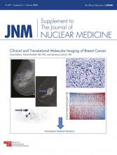
As the supplement editors, it is our pleasure to introduce to you a special supplemental issue of The Journal of Nuclear Medicine on the application of molecular imaging to breast cancer. This issue is part of an ongoing series of JNM supplements focused on diseases for which molecular imaging has a high impact on the approach to clinical care and on translational research. Advances in our understanding of the biologic basis of breast cancer have led to increasingly tailored approaches to breast cancer diagnosis and treatment—a precision medicine approach guided by sophisticated diagnostics and targeted therapeutics. The ability to assess an individual patient’s risk of breast cancer, including genetic predisposition, guides the approach to breast cancer screening and diagnosis. Molecular imaging brings a new dimension to the important tasks of breast cancer diagnosis and staging, and there is considerable potential for novel imaging approaches to molecularly targeted detection. Molecular imaging also provides a unique tool that can guide individualized cancer therapy by assessing therapeutic targets, measuring early response to treatment, and identifying residual viable cancer after therapy. In this supplemental issue, we review the important applications of molecular imaging to breast cancer diagnosis and treatment, highlighting current clinical uses and new approaches under study.
We have divided the issue into an initial overview followed by 3 sections: current clinical applications, emerging applications in diagnosis or as a therapeutic biomarker, and translational research on new approaches and applications. We begin with an overview by Elizabeth McDonald and colleagues (1) of current approaches to breast cancer diagnosis and treatment, highlighting areas in which molecular imaging can play an important role. Next, David Groheux and colleagues (2) review the current use of 18F-FDG PET/CT for breast cancer staging, emphasizing those clinical scenarios in which the modality is most helpful. Gary Cook and colleagues (3) then review the use of 18F-FDG PET/CT and other types of molecular imaging in the important tasks of detecting and evaluating metastases of breast cancer to bone, the most common site of distant disease spread. Norbert Avril (4) finishes the section on current clinical applications by reviewing the use of 18F-FDG PET/CT for measuring breast cancer treatment response in both the neoadjuvant and metastatic settings.
In the section on emerging clinical applications, David Hsu and colleagues (5) and Wendie Berg (6) review the use of molecular imaging for breast cancer detection, focusing on novel instrumentation and current and future clinical uses, respectively. Gary Ulaner and colleagues (7) close the section by providing an overview of the use of 18F-FDG PET/CT and other methods currently under study as imaging biomarkers to help guide individualized treatment of breast cancer patients.
The final and largest section of the issue covers the exciting topics of translational research on new molecular imaging approaches to breast cancer and early testing of their application to breast cancer patients. Charles Manning and colleagues (8) review the preclinical molecular imaging research to elucidate the biology of breast cancer in vivo and to guide clinical applications. Elizabeth McDonald and colleagues (9) describe approaches to the development of new molecular imaging probes—and novel combinations of probes—using examples focused on measurement of breast cancer proliferation/quiescence and DNA damage repair, two rapidly emerging areas of biologic and therapeutic interest. We then include a series of 3 articles focusing on molecular imaging as related to 3 key sets of drug targets and biomarkers. Amy Fowler and colleagues (10) review molecular imaging of steroid receptors in breast cancer, focusing on methods of predicting and measuring the response of the breast to endocrine therapy. Geraldine Gebhart and colleagues (11) discuss methods of imaging expression of HER2 and response to HER2-targeted therapy, highlighting the results of recent clinical trials. Laura Kenny (12) covers imaging methods of measuring proliferation, DNA damage, cell death, and angiogenesis—processes important to breast cancer pathogenesis and progression and key biomarkers of therapeutic response. Finally, Suzanne van Es and colleagues (13) discuss what is required to move a novel molecular imaging method for breast cancer from preclinical testing into early clinical trials and eventually into clinical application.
We conclude the issue with an editorial summary of the field by Richard Wahl (14), an early pioneer in PET imaging of breast cancer, with some thoughts about the current state of the field and how molecular imaging can best serve the needs of breast cancer patients and the physicians who treat them.
In closing, we would like to thank the many authors of this supplemental issue for their efforts and excellent contributions, as well as our reviewers and editorial staff for ensuring a high-quality and comprehensive review of the subject area. We hope that JNM readers will find the issue interesting and helpful and will appreciate the considerable progress in this area along with the tremendous potential for molecular imaging to benefit breast cancer patients.
- © 2016 by the Society of Nuclear Medicine and Molecular Imaging, Inc.
REFERENCES
- Received for publication November 2, 2015.
- Accepted for publication November 3, 2015.


